
Counting the Invisible: How Scientists Track Yeast in Wine Fermentation
By: María Vázquez Fernández (DC2)
09/07/2026
When grape juice begins its transformation into wine, an invisible network of interactions emerges among different yeast species, the key microorganisms responsible for alcoholic fermentation. Millions of yeast cells simultaneously compete, cooperate, and adapt to their environment, ultimately shaping the aromas, flavours, and complexity of the wine.
During this process, not all yeasts act in the same way. While Saccharomyces cerevisiae plays the dominant role in alcoholic fermentation, many other yeast species are present at the early stages of fermentation. These non-Saccharomyces yeasts can significantly influence the process by modifying wine composition in multiple ways, such as reducing ethanol content, modulating acidity, influencing colour stability, impacting aroma, or even causing spoilage (Comitini et al., 2021).
Within the Eco2wine project, understanding which yeasts are present and how their populations change over time due to natural microbial interactions is essential for developing innovative strategies and producing consistent, high-quality wines.
How do scientists determine yeasts are present in wine?
Traditionally, microbiologists have relied on plating techniques, which consist of taking a small sample of fermenting must and spreading it onto nutrient-rich agar plates, where individual yeast cells grow into visible colonies (Wang et al., 2016) (Figure 1). By counting and identifying these colonies, researchers can estimate which species are present and in what proportions. However, this method presents several limitations, such as underestimating certain yeasts, particularly those that grow slowly or fail to grow under laboratory conditions.
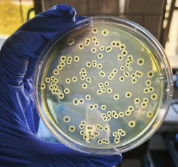
Figure 1. Single colonies of a yeast specie growing on YPD agar plate after incubation.
Nowadays, this technique is being replaced by high-tech alternatives such as flow cytometry. Instead of waiting for colonies to grow, flow cytometry enables the real-time analysis of individual cells. In this approach, cells are analysed one by one they flow through a very narrow channel. This is achieved by a fluidic system that aligns the cells in a single file, allowing them to pass individually through a focused laser beam. As each cell passes through the laser, it interacts with the light in different ways, providing valuable information about cell size, internal complexity, and cell viability (Conacher et al., 2020). Table 1 compares the two techniques, highlighting how and why flow cytometry is a much faster and more reliable alternative, even though it is more expensive.
Table 1. Comparative analysis of counting plates and flow cytometry characteristics.
| Counting Plates | Flow Cytometry | |
| Speed | Slower: Days | Faster: Minutes |
| Detection | May miss some microorganisms | Detect different cell types easily |
| Counting method | Counts colonies grown on plates | Counts individual cells directly |
| Cell viability | Only detects cells that are able to grow | Can distinguish live and dead cells |
| Ease of use | Simple, low-cost, widely used | More expensive, Requires specialised equipment and expertise |
| Limitations | Time-consuming and may underestimate microorganisms | Needs fluorescent dyes and specialised instruments |
How is it possible to discriminate between different yeast species during alcoholic fermentation in winemaking?
Flow cytometry can detect fluorescence signals through highly sensitive detectors that capture different wavelengths emitted by fluorescent molecules. This capability represents a key advantage when working with mixed cultures, as it allows researchers to distinguish and track different subpopulations based on specific fluorescent markers, even when they coexist within the same sample. However, when different yeasts are grown in co-culture, they often display distinct physical and physiological characteristics that are not always sufficient to fully discriminate between all populations. To overcome this limitation, several complementary strategies have been developed to specifically identify and monitor different yeast species during fermentation, many of which rely on fluorescence-based detection within flow cytometry (McKinnon, 2018).
One powerful approach is genetic modification, in which yeast species are engineered to express different fluorescent proteins such as GFP (Green Fluorescent Protein), RFP (Red Fluorescent Protein) or BFP (Blue Fluorescent Protein), among others. In this way, each yeast species is tagged with a different colour, and after being excited with a specific laser, their emission wavelength is captured and processed, allowing the discrimination of different yeast species (Conacher et al., 2020) (Figure 2). Another technique used is Fluorescence In Situ Hybridisation (FISH). This method uses fluorescently labelled DNA or RNA probes that bind to species-specific genetic sequences inside the cells. By targeting unique regions of ribosomal RNA, FISH allows precise identification in mixed cultures (Andorra et al., 2011). In addition, fluorescent dyes provide valuable information by revealing different physiological characteristics of yeast cells. Among these, the most commonly used in mixed cultures are viability stains, such as Propidium iodide, cFDA or SYTO9 (Sommer, 2020).
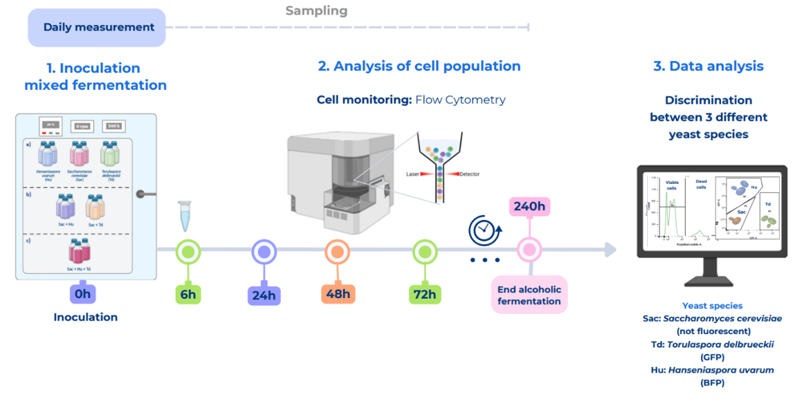
Figure 2. Experimental design for the discrimination of three different yest species during alcoholic fermentation using flow cytometry. Sac: Saccharomyces cerevisiae (unlabelled), Td: Torulaspora debrueckii (GFP-labeled) and Hu: Hansenisapora uvarum (BFP-labeled). Daily measurements are performed to monitor the viability of each species based on their specific fluorescence signals.
Flow cytometry is a powerful tool for understanding yeast dynamics, as it provides detailed, real-time and highly informative data. This enables finer control over the fermentation process, allowing researchers and winemakers to optimise fermentations, limit the growth of undesirable microorganisms, and better guide the development of aromas and flavours. As this technology continues to advance, it is paving the way for greater control, deeper understanding, and increased innovation in winemaking, effectively bridging the gap between microbiology and sensory experience.
Bibliography
Andorra, I., Monteiro, M., Esteve-Zarzoso, B., Albergaria, H., & Mas, A. (2011). Analysis and direct quantification of Saccharomyces cerevisiae and Hanseniaspora guilliermondii populations during alcoholic fermentation by fluorescence in situ hybridization, flow cytometry and quantitative PCR. Food microbiology, 28(8), 1483-1491. https://doi.org/10.1016/j.fm.2011.08.009
Comitini, F., Agarbati, A., Canonico, L., & Ciani, M. (2021). Yeast interactions and molecular mechanisms in wine fermentation: a comprehensive review. International Journal of Molecular Sciences, 22(14), 7754. https://doi.org/10.3390/ijms22147754
Conacher, C. G., Naidoo-Blassoples, R. K., Rossouw, D., & Bauer, F. F. (2020). Real-time monitoring of population dynamics and physical interactions in a synthetic yeast ecosystem by use of multicolour flow cytometry. Applied microbiology and biotechnology, 104(12), 5547-5562. https://doi.org/10.3109/07388551.2015.1128876
McKinnon, K. M. (2018). Flow cytometry: an overview. Current protocols in immunology, 120(1), 5-1. https://doi.org/10.1002/cpim.40
Sommer, S. (2020). Monitoring the functionality and stress response of yeast cells using flow cytometry. Microorganisms, 8(4), 619. https://doi.org/10.3390/microorganisms8040619
Wang, C., Mas, A., & Esteve-Zarzoso, B. (2016). The interaction between Saccharomyces cerevisiae and non-Saccharomyces yeast during alcoholic fermentation is species and strain specific. Frontiers in microbiology, 7, 502. https://doi.org/10.3389/fmicb.2016.00502